App Note I May 17, 2017
List of protein hydrodynamic diameters DH
| Protein | Molecular weight | DH measured with switchSENSE® (i) | Dh calculated from PDB file (ii) | PDB code | Structure |
|---|---|---|---|---|---|
| anti-human Fab fragment | 50 kDa | 6.6 nm | 7.2 nm | 3WD5 |  |
| anti-human IgG | 150 kDa | 12.7 nm | 11.2 nm | 1IGT | 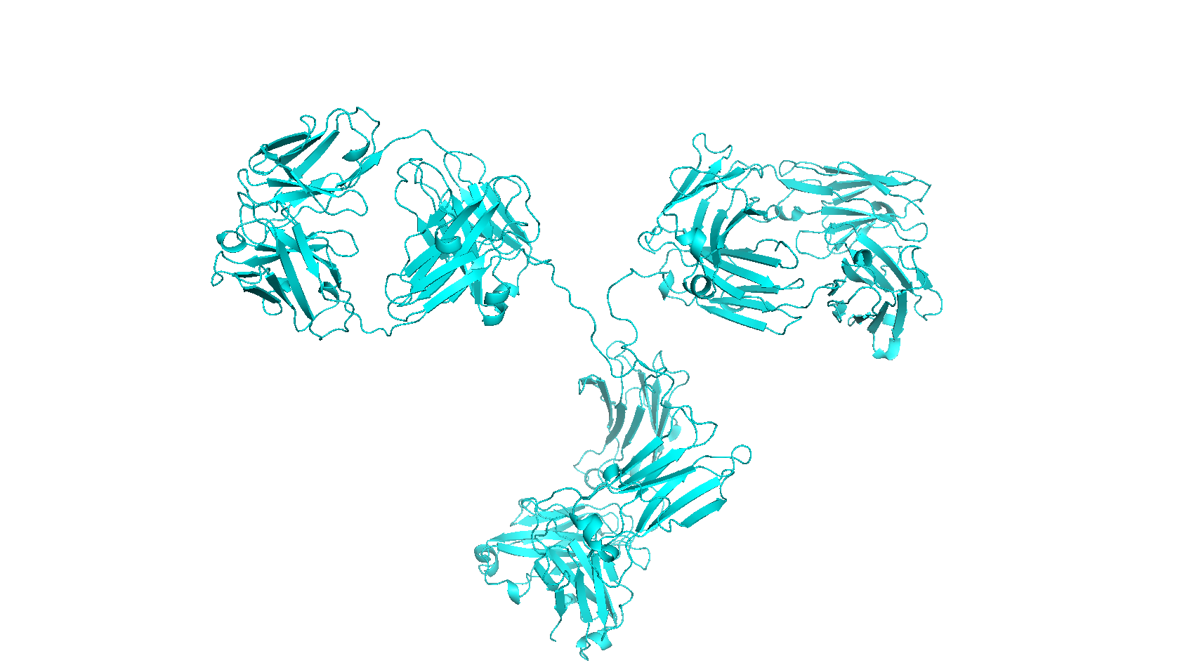 |
| anti-IgG Affibody® | 14 kDa | 3.9 nm | 3.9 nm | 2B88 | 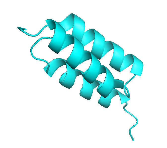 |
| Carbonic anhydrase I | 29 kDa | 4.9 nm | 4.9 nm | 2CAB | 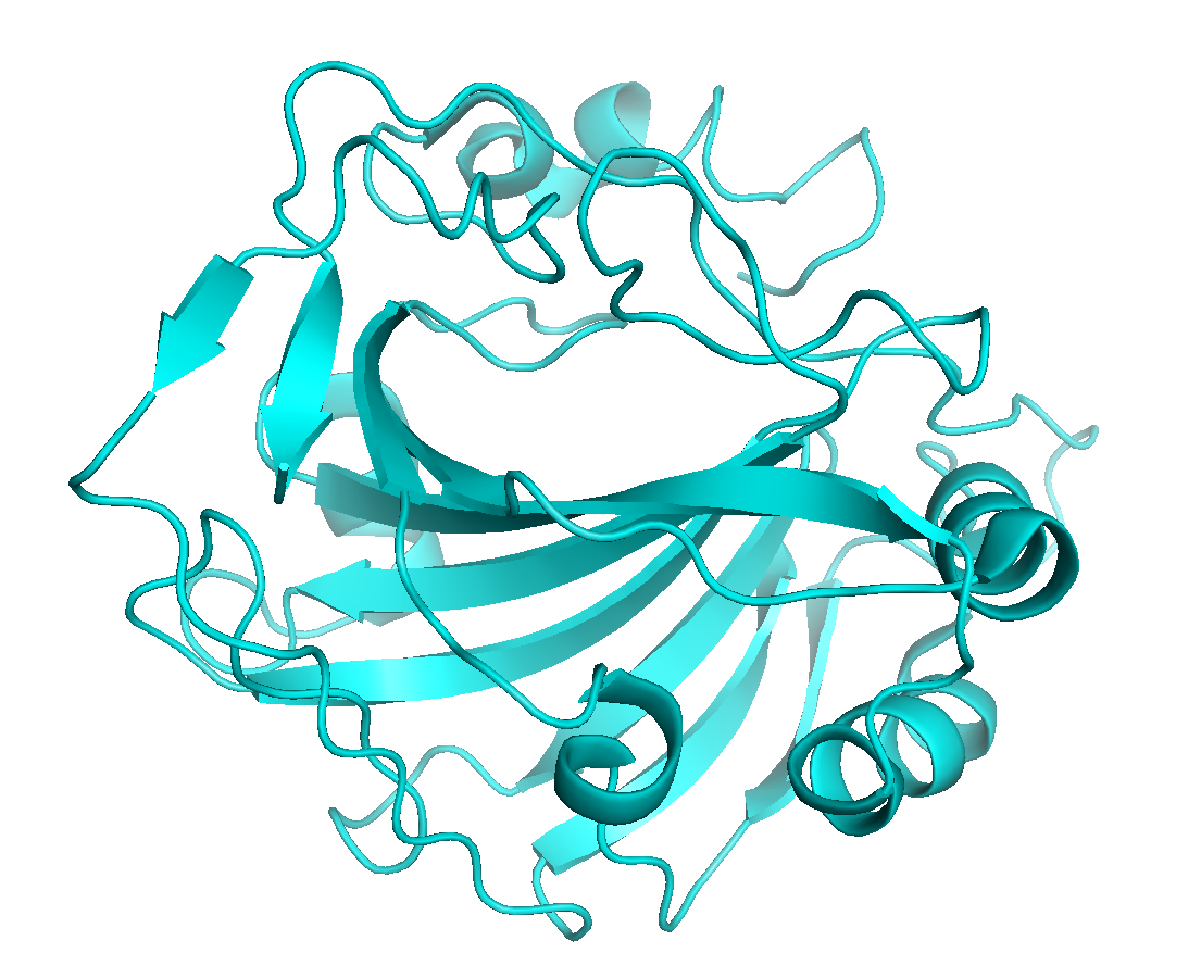 |
| CD64 | 31 kDa | 6.7 nm | 5.7 nm | 4ZNE | 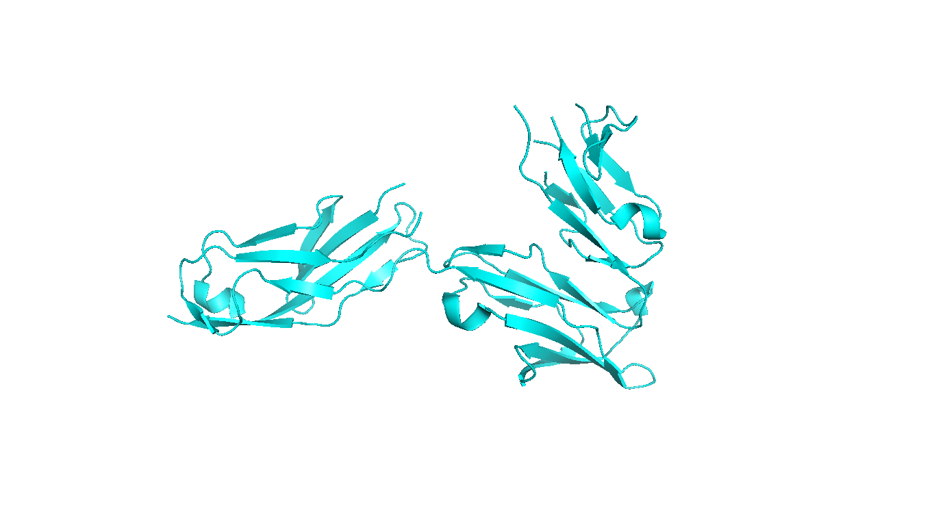 |
| EGFR | 71 kDa | 7.1 nm | 7.8 nm | 1NQL | 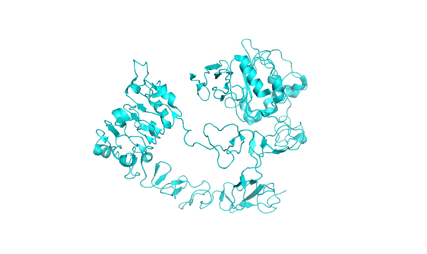 |
| Erythropoetin | 21 kDa | 4.7 nm | 4.6 nm | 1BUY | 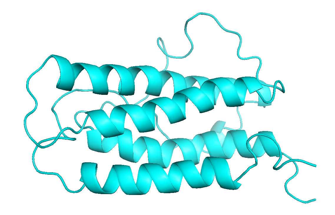 |
| GFP-Trap® (nanobody) | 13 kDa | 3.9 nm | 3.9 nm | 3OGO | 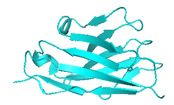 |
| Glutathion-S-Transferase | 26 kDa | 4.7 nm | 4.8 nm | 1B8X | 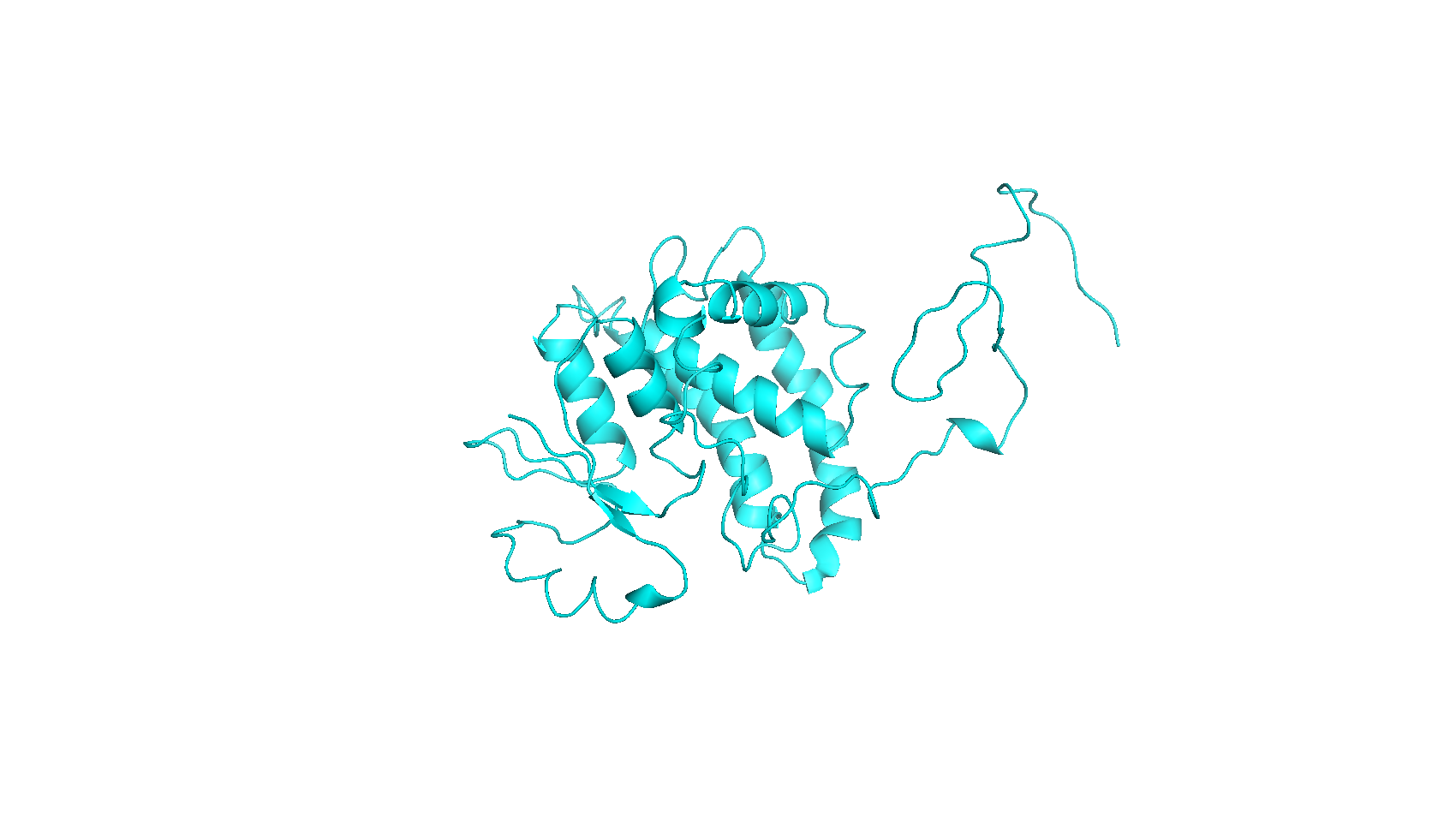 |
| Glycosylceramidase | 56 kDa | 6.4 nm | 6.2 nm | 3RIL |  |
| Her2 (Met1-Thr652) | 70 kDa | 7.6 nm | 7.7 nm | 1N8Z | 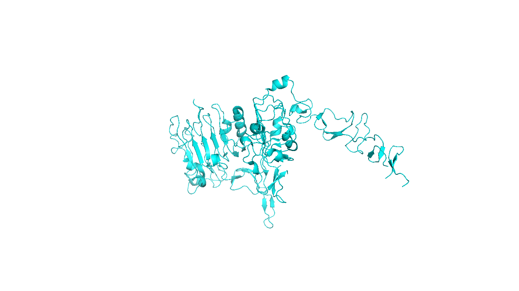 |
| HIV Env protein | 500 kDa | 11.0 nm | 11.5 nm | 4NCO |  |
| Human serum albumin | 69 kDa | 4.2 nm | - | 1AO6 | 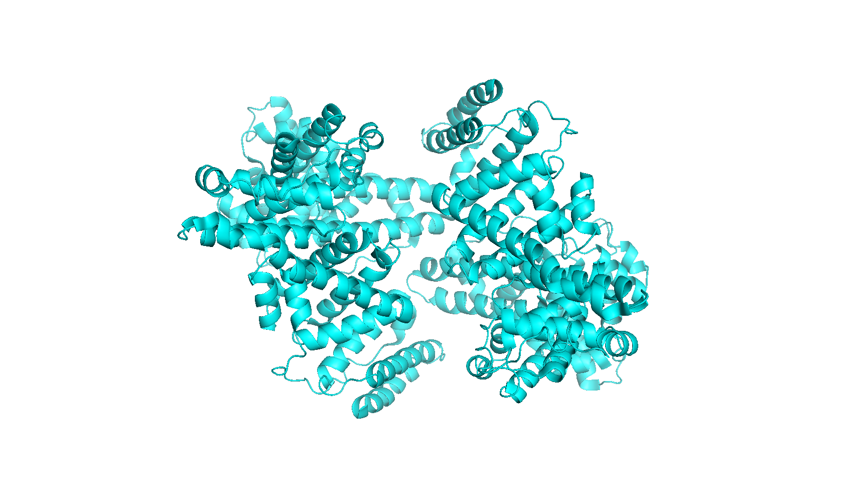 |
| Neutravidin | 60 kDa | 7.4 nm | 6.1 nm | 1VYO | 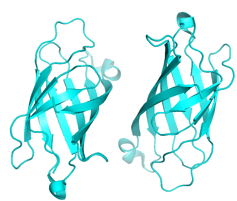 |
| p38 kinase | 41.5 kDa | 5.0 nm | 5.8 nm | 1W84 | 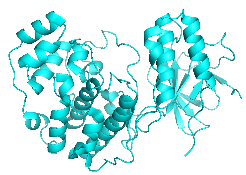 |
| Pim-1 | 31.5 kDa | 5.2 nm | 5.2 nm | 3C4E | 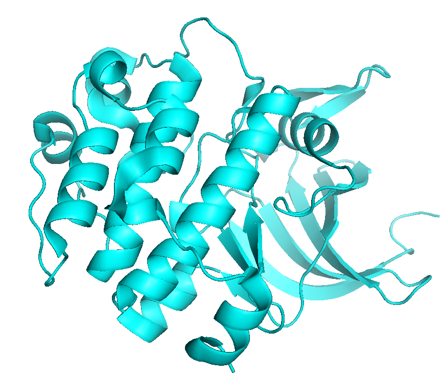 |
| Protein kinase A | 40 kDa | 5.5 nm | 5.6 nm | 1Q8U | 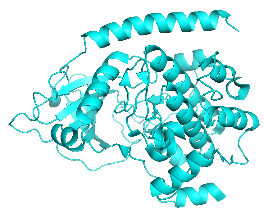 |
| Pyruvate kinase M2 | 58 kDa | 6.9 nm | 6.4 nm | 1T5A | 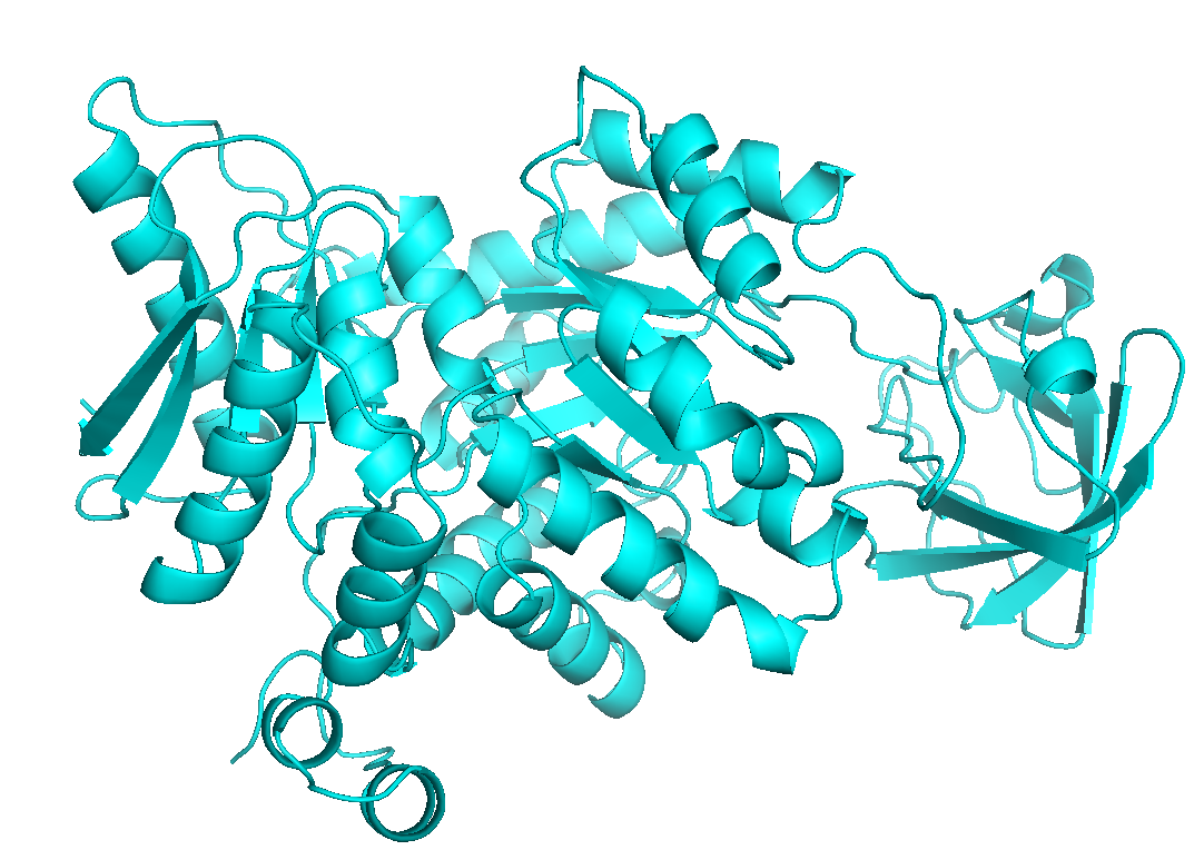 |
| Sting dimer | 56 kDa | 6.5 nm | 6.6 nm | 4F5D |  |
| Sting monomer | 28 kDa | 5.1 nm | 4.9 nm | 4F5E | 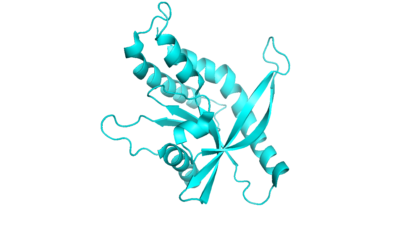 |
| TNFα | 17.5 kDa | 4.4 nm | 4.4 nm | 3WD5 | 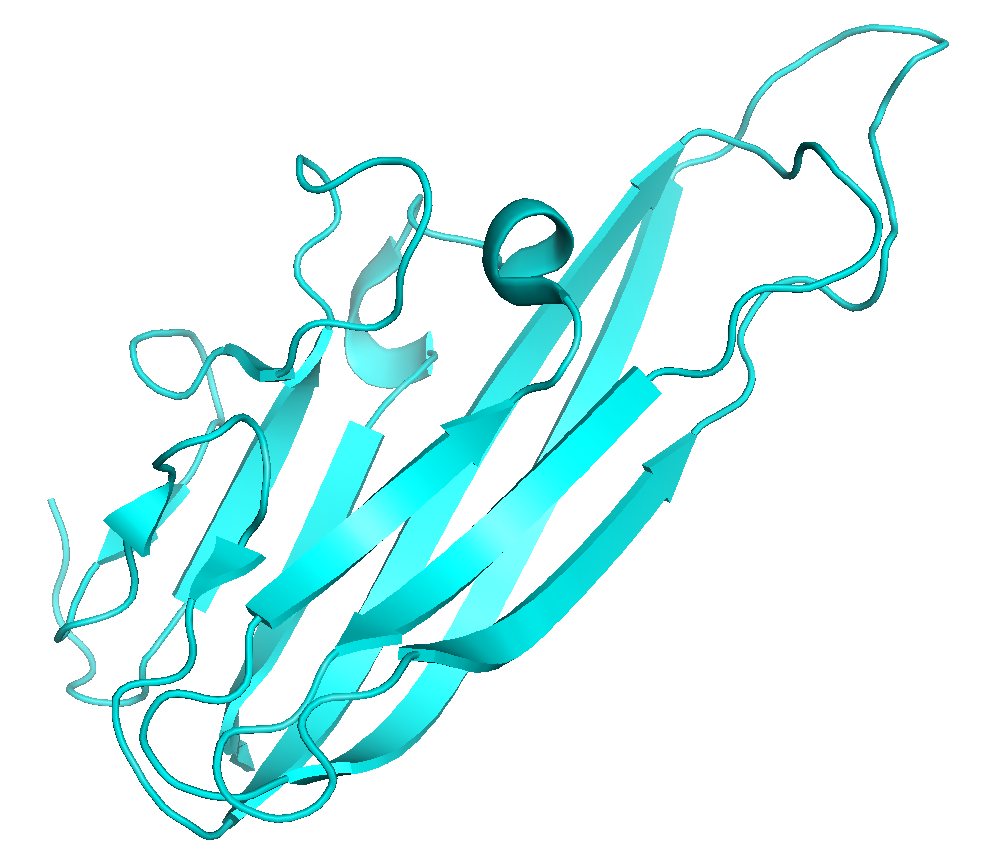 |
| Ubiquitin | 8.5 kDa | 3.2 nm | 3.4 nm | 1UBQ | 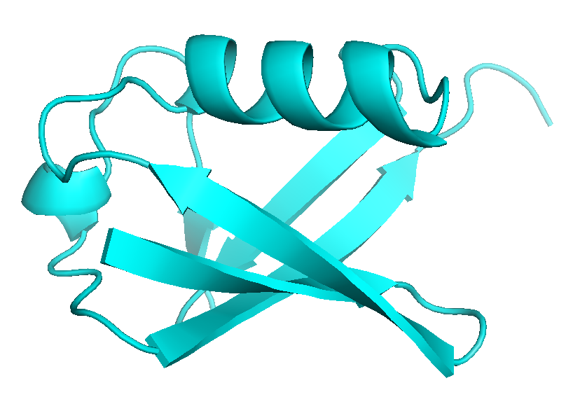 |
(i) Experimental protein diameters are analyzed from switching dynamics measurements with standard 48 bp DNA nanolevers (length = 16 nm) using the drift-diffusion ‘Lollipop’ model described in J. Phys. Chem. B 118, 597 (2014).
(ii) Theoretical calculations of protein diameters from PDB files are performed with the HydroPro algorithm [Garcia de la Torre et al. Biophys. J. 78:179, (2000)], that calculates an effective hydrodynamic diameter from atomic coordinates using a hydrodynamic bead-shell model. Note that crystal structures may not always reflect the native protein structure in solution (especially for large proteins with several domains connected by flexible linkers).